Double Trouble: 2 Circulatory Systems and 2 Hearts

Tags

, , , , , ,

Whether we crawl, run, or swim, we all depend on our circulatory systems to transport oxygen, carbon dioxide and nutrients to the appropriate tissues while excreting metabolic waste; regulate hormones and temperature; and protect us against foreign agent through immune function.

There are two types of circulatory system.

The first type is the Open Circulatory System. It consists of a primitive heart that pumps hemolymph (fluid analogous to blood) into the hemocoel (body cavity that contains fluids). With these structures in place, the blood pressure is low and the blood volume is relatively large, constituting 20-40% of body volume. It is almost impossible to directly control how fast the blood spreads through the body, as the flow of blood is dependent on body movement.

figure-12-02a

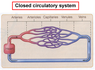
In the Closed Circulatory Systems, blood flows in a continuous circuit where it is exposed to high pressures that allow for high oxygen uptake and blood volume only consists of 5-10% body volume. The system relies on all components to ensure blood flow. The components are as follows:

  • propulsive organ (heart): forces blood through the body
    • capillaries:  a network of miniature blood vessels that facilitates gas exchange
  • arterial system: series of blood vessels that are surrounded by a lot of muscle that allows blood to be propelled from the heart and distributed throughout the body.
      • arteries: only hold 16-18% of blood volume
  • venous system: blood vessels that carry deoxygenated blood from the blood back to heart. Veins are much more elastic and can expand quite easily to hold more volume
  • veins: can hold up to 50 % of blood

Screen Shot 2015-04-20 at 6.58.43 PM

Note: The distribution of blood in the circulatory system is as follows:

  • Pulmonary circulation is responsible for 12% of blood flow
  • The heart only contains 15 % at a time
  • The capillaries and arterioles hold the least about of 5% – 7%
  • While the arteries carry 16% – 18% at a time
  • The veins and venules have the capacity to hold up to 50%

In Closed Circulatory Systems, there are also 2 types of hearts. First we will look at the neurogenic heart which depends on nerves to maintain rhythm, and then we will take a look at the myogenic heart that consists of muscle which generates the beating of the heart.


Screen Shot 2015-04-20 at 6.57.19 PM
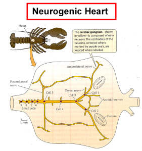
The image above displays a neurogenic heart, which is generally found in anthropods (an invertebrate animal with an external skeleton segmented body and jointed appendages) and officially referred to as the crustacean cardiac ganglion.  The small cells (9,8,7,6) act as the pacemaker that conducts electrical impulses to stimulate contractions. This heart can be found in insects, crayfish, crab or lobsters.


heart_disease_-_general-2
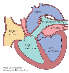
The myogenic heart has 4 chambers, left and right atria and left and right ventricles. The sinoatrial node located in the wall of the right atrium acts as a pacemaker, where cells depolarize spontaneously to control the beating of the heart. The blood in the atria is pumped through valves into the ventricles that supply the main propulsive force to distribute blood through the body.

Saturday Night Fever

Tags

, , , , , ,

Feverish-Feeling-With-Chills-300x300    Feeling feverish?

Ever wonder how fevers happen?

cute_lizard
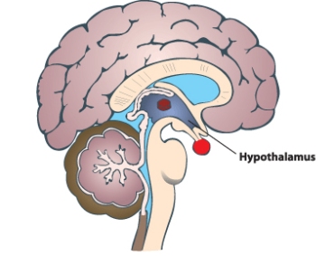
Or maybe your pet iguana has a fever?


For some this may sound strange, but ectotherms (cold-blooded animals) also get fevers, which cause them to bask in radiant heat zones for longer periods of time.


A fever is a phenomena where body temperatures are elevated due to an immune response in which pyrogens are released in response to infection and/or inflammation. The pyrogens that are released from into the bloodstream find their way to the brain and bind to neurons in the hypothalamic thermoregulatory center to alter the information received. Pyrogens disrupt neuronal activity causing the warm sensitive neuronal activity to be reduced and the cold sensitive neuronal activity to increase. The hypothalamus region of the brain perceives that the body is cold and increases heat production which leads to an elevation of the body’s set temperature because of the perceived cold temperatures.

Let’s take a closer look at the physiology behind temperature regulation to better understand fevers.

Peripheral thermal receptors on the skin, and central receptors in parts of the brain and abdominal region send messages about changes in body temperature to the “thermostat” known as the hypothalamus (a small portion of the brain that regulates body temperature).

Hypothalamus

Upon receipt of this information the hypothalamus makes the adjustment by altering activity in three neuronal populations.

Neuron population 1 – cold sensitive:

  • generates heat via vasoconstriction (narrowing of blood vessels) and piloerection (an involuntary action that cause the hair to stand upright)

Neuron population 2 – warm sensitive:

  • dissipates heat via vasodilation (broadening of blood vessels) and sweating

Neuron population 3  – warm sensitive:

  • reduces thermogenesis (production of heat) by minimizing vasoconstriction and eliminating piloerection.

Note:

Vasoconstriction maintains body temperature and reduces heat loss by constricting blood vessels in which blood flow to the skin is reduced. Blood cools when close to the surface of skin.  During vasodilation, blood vessels are broadened and the blood vessels rise to the skin’s surface, allowing the blood to dissipate heat from the body.

Similarly, in response to colder temperatures piloerection generates heat when the hairs stand in an erect position forming an insulating layer that traps heat.  Whereas, in hot conditions, the hairs remain flat to prevent the insulating layer from developing.

The significance of fevers have not been identified, but there are speculations that fevers improve recovery time. A 1994 study of fever suppression in children with chickenpox showed that the recovery time in children that took anti-fever medication was much longer than those who allowed the fever to take its course.

My goal of sharing this information is to inform you of the physiology of fevers so that you may relax, feeling reassured with the knowledge obtained. Thanks for reading!

Heat Shock Proteins – the ultimate stress fighters

Tags

, , , ,

Deep thought

Every once in a while, youtube break provides that quick entertainment needed to generate ideas and recharge. While studying for my physiology exam, I came across a video on Heat Shock Proteins (HSPs) by @ChrisGoneRaw claiming that HSPs can accelerate post-workout recovery time.

I was absolutely stunned by this claim.

Could it be true?

Can HSPs really help me recover sooner from a hard workout?

This lead me to ask myself, well what are these HSPs anyways? To answer this question I had to consult my lecture notes and an article to ensure I had all the facts.

What are heat shock proteins (HSPs)?

Heat-shock proteins (HSPs) are a family of thermostable, hydrophobic proteins that are produced  by heat-shock cells in response to environmental stress. Heat shock cells are highly conserved, meaning they appears in every organisms across generations.

HSPs were first discovered in the 1960’s by an Italian geneticist who observed the increase synthesis of proteins in the cells of fruit flies when exposed to heat stress. Now, with further investigation over the years, research revealed that HSPs respond to various forms of stress including exposure to excessive heat, cold, ultraviolet light, and oxidative stress; even wound healing can induce the expression of HSPs.

HSPs consist of a large family of proteins that are classified based on molecular weight which are measured in kilodaltons (kD).

  • hsp10 = 10 kD
  • hsp40 = 40 kD
  • hsp60 = 60 kD
  • hsp70 = 70 kD
  • hsp90 = 90 kD

There are two forms in which HSPs are expressed. First, as inducible proteins that appear during stress, and as molecular chaperones that are always present. The molecular chaperones play various roles which you will see listed below.

Inducible proteins:

  • only expressed when induced by stress

Molecular chaperones:

  • always present
  • maintains cell-cycle control and signaling
  • protects cells against stress and cell death (apoptosis)
  • refolds denatured proteins that have been exposed to stress
  • assist with protein the folding of newly formed polypeptides produced by ribosomes
  • transports and sorts proteins into appropriate subcellular compartments

Now that we know what HSP proteins are, let’s take a look at how they work.

Courtesy of Dr. Spencer Mukai

Courtesy of Dr. Spencer Mukai

In a stress-free environment, HSPs are bound to monomer (individual units) of heat shock factor (HSF-1), forming a folded protein complex. However, the moment stress arises, the protein complex containing HSPs and HSF-1 unfold, allowing the HSF-1 monomer to unite with two other monomers to form a HSF-1 trimer. The trimer enters the nucleus via translocation (moving a portion of a chromosome to a new position). Once inside the nucleus the HSF-1 trimer binds to the heat shock enhancer (HSE)  in the promoter region; activating the transcription of HSP mRNA,  which will be converted to HSPs. In other words more HSPs are produced to respond to stress within the body.


So after all this, it turns out that @ChrisGoneRaw’s claim that HSPs can accelerate post-recovery was correct! As demonstrated in the photo above, if stress is present in the cells, the HSPs-HSF-1 units will separate triggering an upregulation of heat shock proteins. The next you find yourself in extreme temperatures, or exposed to ultraviolet and/or oxidative stress (for short periods of time), remember that the heat shock proteins in your body are protecting your cells so help fight against stress.

Wart-ever it’s just Hot Yoga: An Overview of Plantar Warts

Tags

, ,

In preparation for the 12 PM yoga session; I relaxed in Savasana ready to start.Planters Warts

Ready to relieve stress and find serenity in my breathing.  Just looking forward to an hour of invigorating and energizing my body. As we transitioned into Vinyasa, sychronizing our breath with our movement. I felt so alive and inspired to attain my yoga goals; until suddenly…

We transition into downward facing dog, and there it was!

A thick, hardened, circular patch of skin with dark spots in the middle of it, appeared on the ball of the foot of the fellow yogi in front of me. This my dear blog followers, was a Plantar Warts. A contagious, and painful skin growth caused by the human papilomavirus (HPV) infecting the skin through a cut or skin abrasion.

As you may or may not know, Warts can be transferred from person to person or through indirect contact.  If the HPV virus strains specific to Plantar Warts are stored in a formite such as a towel or a moist yoga mat; then this exposure would increase the likelihood of transmitting the virus and spreading warts.  Individuals with compromised immune systems are even more susceptible to contracting warts.

Fortunately there are a number of treatments for Plantar Warts.

Salicylic acid can be used to softens the skin that forms the wart so that it can be easily rubbed off.  Compound W has been effective.

Over the counter Cryotherapy  where liquid nitrogen is applied to the wart to freeze  and remove it. Dr. Scholl’s Freeze Away is available without a prescription.

Retinoid creams are derived from vitamin A  and have be proven to be an effective treatment for common warts.

A visit to the doctor’s office for Cantharidin is also an alternative treatment. It is injected into the wart causing the skin under the wart to blister, and lift the wart off the skin.

Treatments for more difficult warts include:

Electrosurgery– the use of an electrical current to burn warts.

Curettage– cutting the wart off with a sharp knife and scooping the tissue for wart removal.

Note: Electrosurgery and Curettage are often used together to treat warts.

Keep in mind that treatment may be painful. The black dots seen in the middle of the warts are tiny blood vessels that grow to supply it with blood.

Although there are several treatment options available, maintaining good hygiene is important for prevention.  Minor cuts should be treated, contact with a wart on another person’s skin should be evaded and hands must be washed carefully after touching a wart.

With these steps Plantar warts can be controlled; however it is most important to see a health care professional if you are unsure if the skin growth is a wart. Lastly, never attempt to cut or burn off a wart by yourself.

To learn more about Planter Warts, you can visit http://www.ncbi.nlm.nih.gov/pubmedhealth/PMH0001888/

Source: Warts. (2012). A.D.A.M Medical Encyclopedia. Retrieved from http://www.ncbi.nlm.nih.gov/pubmedhealth/PMH0001888/